


Animal cell cutaway: structure in three dimensions
Inspect a nucleated animal-cell schematic with membranes, organelles and contrasting transport compartments. The open geometry makes selected relationships visible from several angles.
Inside the assembly
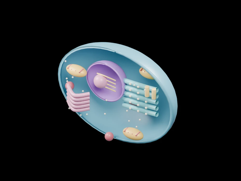
A retained blue outer wall creates a front opening. The purple nuclear shell contains a pink nucleolus and gold chromatin guides. On one side, repeated teal membrane paths and pale dots represent rough ER with ribosomes. A separate pink stack forms the Golgi illustration. Orange organelles with short inner strokes indicate mitochondria; smaller coloured spheres distinguish additional compartments.
Scientific explanation
Membranes organise a eukaryotic cell into compartments. The nucleus encloses chromosomes, while pores support controlled exchange with the cytoplasm. Ribosomes, membrane networks and other organelles support different cellular processes.
- Nuclear envelope
- Membrane boundary around the nucleus
- ER
- Endoplasmic-reticulum membrane network
- Golgi apparatus
- Compartment involved in processing and trafficking
Organelle numbers, sizes and locations are illustrative. Chromatin is not a DNA sequence, and no specific cell type, biochemical kinetics or live-cell state is represented. Colours and proportions are teaching conventions. This original schematic is not patient-derived or clinically validated, and is not a molecular simulation or a manufacturing specification.
Explore the model
- Find the nuclear opening and the smaller nucleolus.
- Distinguish dotted rough ER from the separate Golgi stack.
- Locate the orange mitochondria and smaller lysosome symbols.
From geometry to engineering
Rotate the model, compare its named structures with the references, then use the article to distinguish what is represented from what is simplified.