


Tailed bacteriophage: structure in three dimensions
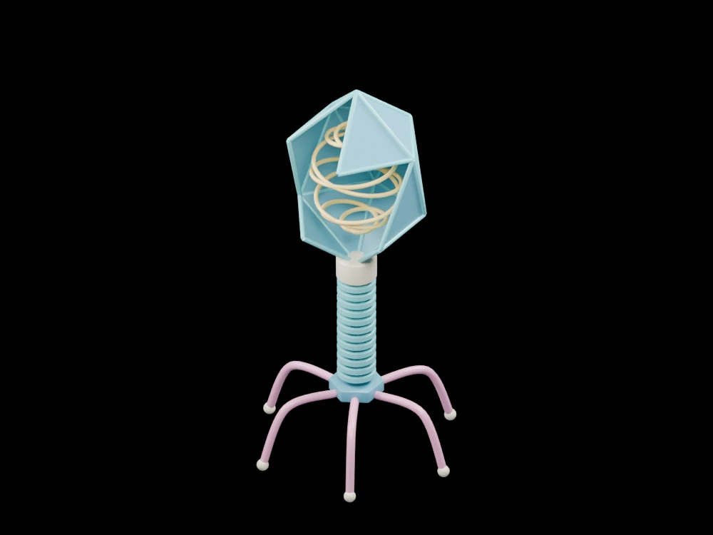
Explore a faceted capsid, packaged-genome illustration, tail sheath, baseplate and six attachment fibres. The open geometry makes selected relationships visible from several angles.
Inside the assembly
An open faceted head exposes a coiled gold line. The neck leads into a thin tail tube surrounded by a stack of sheath segments. A hexagonal plate at the lower end supports six outward-bending fibres. The edge rods emphasise the capsid outline and are not an additional biological layer. Every part is original teaching geometry, with no atomic coordinates or experimental viral data.
Scientific explanation
Bacteriophages are viruses that infect bacteria. Tailed forms have a protein head containing genetic material and attachment/delivery structures. Viral shapes vary widely; this model illustrates one general tailed arrangement rather than all phages.
- Capsid
- Protein enclosure around genetic material
- Tail sheath
- Illustrated surrounding tail structure
- Tail fibres
- Structures associated with host attachment
This is a nonfunctional structural illustration, not a specified viral strain, genome, host-recognition model or experimental protocol. Head symmetry and dimensions are simplified. Colours and proportions are teaching conventions. This original schematic is not patient-derived or clinically validated, and is not a molecular simulation or a manufacturing specification.
References
OpenStax: viral morphologyExplore the model
- Look through the capsid opening at the coiled genome symbol.
- Distinguish the narrow central tube from the sheath rings.
- Count the six long fibres extending from the baseplate.
From geometry to engineering
Rotate the model, compare its named structures with the references, then use the article to distinguish what is represented from what is simplified.