


DNA double helix: structure in three dimensions
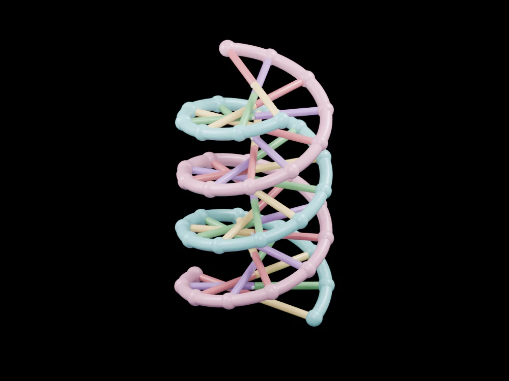
Compare two helical backbones and complementary base-pair symbols in a short enlarged DNA segment. The open geometry makes selected relationships visible from several angles.
Inside the assembly
Two differently coloured backbone paths wind around a shared axis. Rounded groups mark their repeated units. At each level, a two-colour connector represents a complementary base pair; small central dots are bonding symbols rather than complete atom positions. The display contains twenty-one base-pair levels and roughly two turns, with regular spacing chosen to make the paired organisation easy to inspect.
Scientific explanation
DNA consists of linked strands with sugar–phosphate backbones and complementary bases. Adenine pairs with thymine; guanine pairs with cytosine. The double-helical arrangement organises this paired information-bearing structure.
- Backbone
- Alternating sugar and phosphate groups
- Base pair
- A complementary A–T or G–C association
- Helix
- A strand winding around an axis
Base letters identify teaching pairs, not a functional genetic sequence. Grooves, atomistic bonding, hydration, exact nucleotide geometry and sequence-dependent structure are omitted. Colours and proportions are teaching conventions. This original schematic is not patient-derived or clinically validated, and is not a molecular simulation or a manufacturing specification.
References
NHGRI: double helixExplore the model
- Follow one backbone without switching colours.
- Compare the alternating paired connector colours.
- Rotate along the axis to see the twist rather than a flat ladder.
From geometry to engineering
Rotate the model, compare its named structures with the references, then use the article to distinguish what is represented from what is simplified.