


Eye optical cutaway: structure in three dimensions
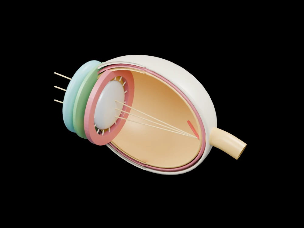
Inspect the cornea, iris, lens, layered wall and optic nerve along an open optical pathway. The open geometry makes selected relationships visible from several angles.
Inside the assembly
Three nested retained shells expose the wall layers. The coloured annular iris has a central opening, with a pale flattened lens behind it and a ring around the lens for the ciliary region. Fine radial strands indicate its suspension. Gold lines illustrate a possible incoming-light route through the open centre; they are guides rather than calculated rays. A thicker yellow extension marks the optic nerve.
Scientific explanation
The cornea and lens contribute to focusing light on the retina. The pupil is the opening controlled by the iris. Retinal photoreceptors convert light into signals that travel towards the brain through the optic nerve.
- Pupil
- Opening within the iris
- Retina
- Light-sensitive tissue at the back of the eye
- Optic nerve
- Route carrying retinal signals
Opaque teaching colours replace transparent tissues. Rays are not an optical calculation, and this model does not support diagnosis, lens prescription or surgical planning. Colours and proportions are teaching conventions. This original schematic is not patient-derived or clinically validated, and is not a molecular simulation or a manufacturing specification.
References
National Eye Institute: how eyes workExplore the model
- Find the pupil opening inside the green iris.
- Compare the nested wall surfaces at the cut edge.
- Trace the illustrative light path and locate the separate optic nerve.
From geometry to engineering
Rotate the model, compare its named structures with the references, then use the article to distinguish what is represented from what is simplified.