

Four-chamber heart: structure in three dimensions
Explore four open chambers, the septum, valve references and the main vessels of a schematic human heart. The open geometry makes selected relationships visible from several angles.
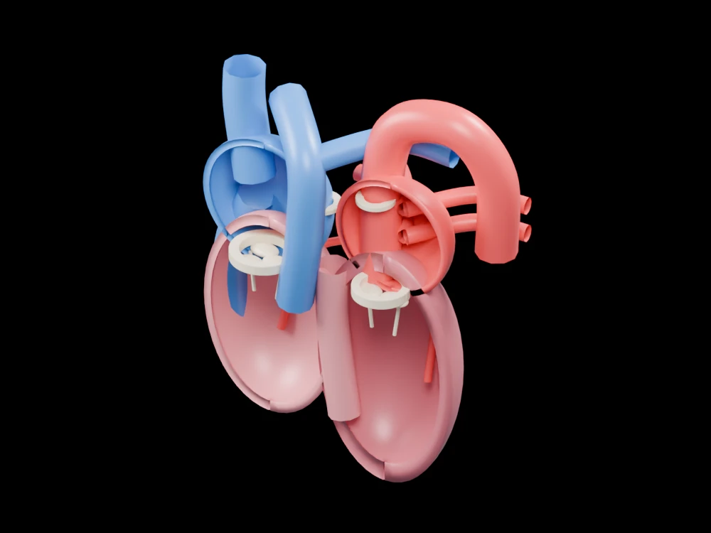
Inside the assembly
Four retained chamber walls form an open front. The anatomical right appears on the viewer’s left in the initial view. Pale rings locate the atrioventricular valves; thinner chordal strands extend below them. The arch above the left side marks the aortic route, while four separate pulmonary-vein branches approach the left atrium behind the chamber walls. The cut surfaces and colour contrast are deliberate viewing aids.
Scientific explanation
The atria receive returning blood and the ventricles pump it onward. The right heart sends blood towards the lungs; the left heart sends it towards the body. Valves limit reverse flow between successive compartments.
- Atria
- The two receiving chambers
- Ventricles
- The two pumping chambers
- Septum
- Tissue separating right and left sides
Chamber orientation, wall thickness and vessel routes are simplified; the model does not simulate pressure, flow, valve motion or a specific heart. Colours and proportions are teaching conventions. This original schematic is not patient-derived or clinically validated, and is not a molecular simulation or a manufacturing specification.
Explore the model
- Find the chamber on each side of the septum.
- Trace the aortic arch and distinguish it from the pulmonary trunk.
- Identify the four pulmonary-vein teaching branches behind the left atrium.
From geometry to engineering
Rotate the model, compare its named structures with the references, then use the article to distinguish what is represented from what is simplified.