


Knee joint assembly: structure in three dimensions
Explore the distal femur, tibial plateau, patella, menisci and selected stabilising ligaments. The open geometry makes selected relationships visible from several angles.
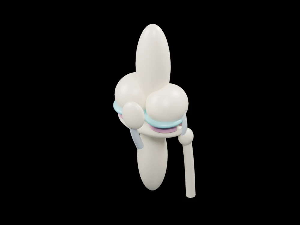
Inside the assembly
Two rounded femoral condyles sit above separated cartilage caps and pink meniscal crescents. The tibial plateau is supported by a lower shaft, with a slimmer fibula alongside it. The patella is positioned anteriorly to expose the central ligament paths. Two crossing strands indicate cruciate attachments, while side strands mark collateral support. Parts are separated slightly to keep the teaching view readable.
Scientific explanation
Articular cartilage helps opposing bone surfaces move smoothly. The two menisci distribute contact and support stability. Ligaments join bones and constrain motion, while the patellar tendon connects the patella to the tibia.
- Menisci
- Two fibrocartilage structures between femur and tibia
- Cruciate ligaments
- Crossing stabilising ligaments inside the joint
- Patella
- Bone positioned at the front of the knee
Attachment positions, articular geometry and tissue thickness are schematic. No ligament forces, joint contact, injury mechanics or range of motion are calculated. Colours and proportions are teaching conventions. This original schematic is not patient-derived or clinically validated, and is not a molecular simulation or a manufacturing specification.
References
AAOS: normal knee anatomyExplore the model
- Identify the two pink crescents above the tibial plateau.
- Rotate to distinguish the anterior patella from the central ligaments.
- Compare the thick tibial shaft with the slim adjacent fibula.
From geometry to engineering
Rotate the model, compare its named structures with the references, then use the article to distinguish what is represented from what is simplified.