


Mitochondrion cutaway: structure in three dimensions
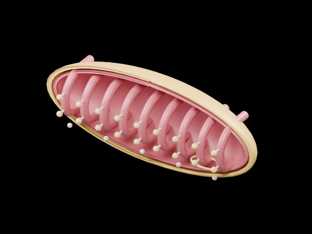
Inspect nested membranes, exposed cristae and schematic ATP-synthase structures within an enlarged mitochondrion. The open geometry makes selected relationships visible from several angles.
Inside the assembly
The outer gold shell and smaller red inner boundary are opened towards the viewer. Repeated red membrane folds extend across the interior; small pale stalks and gold heads indicate ATP-synthase locations without recreating a protein structure. A gold loop and pale particles mark mitochondrial-DNA and ribosome concepts within the matrix. Rotating the view separates the outer envelope from the folded inner surface.
Scientific explanation
Mitochondria have outer and inner membranes. Electron transport across the inner membrane helps establish a proton gradient. Proton flow through ATP synthase is coupled to ATP formation; the folded membrane provides space for these processes.
- Cristae
- Folds of the inner mitochondrial membrane
- Matrix
- Compartment enclosed by the inner membrane
- ATP synthase
- Enzyme coupling proton flow to ATP production
The nine folds and enzyme count are illustrative. No atomistic protein structure, membrane potential, respiration rate or ATP yield is calculated. Colours and proportions are teaching conventions. This original schematic is not patient-derived or clinically validated, and is not a molecular simulation or a manufacturing specification.
Explore the model
- Distinguish the outer shell from the folded inner membrane.
- Locate the enlarged stalk-and-head enzyme symbols.
- Find the circular DNA teaching loop in the matrix region.
From geometry to engineering
Rotate the model, compare its named structures with the references, then use the article to distinguish what is represented from what is simplified.