

Vertebral column: structure in three dimensions
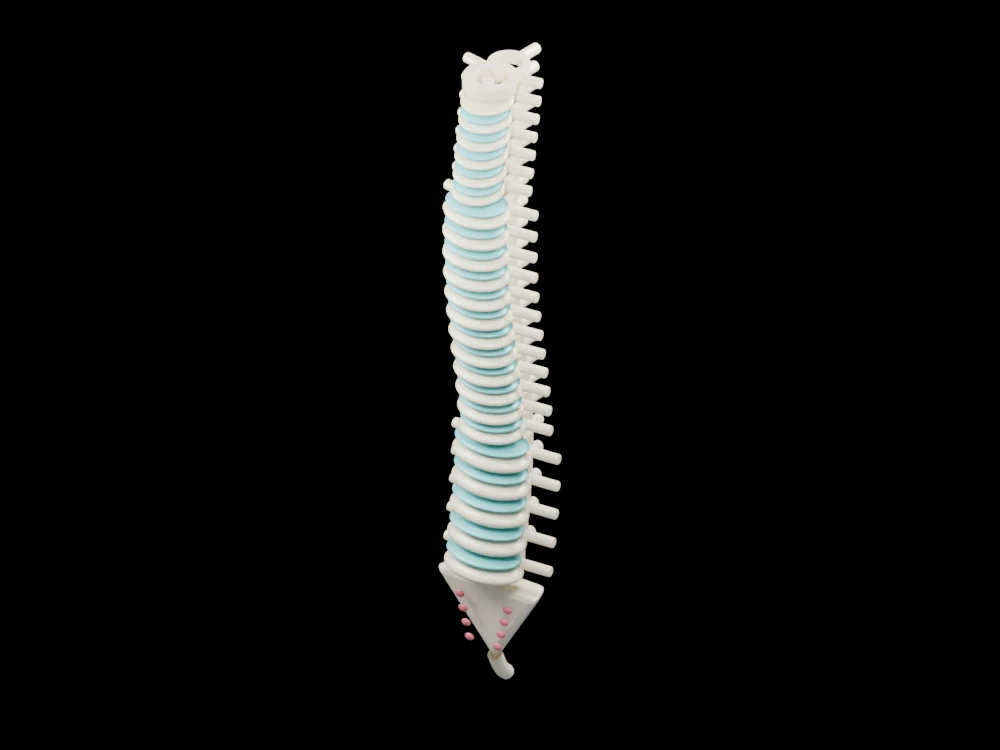
Compare cervical, thoracic and lumbar regions above simplified fused sacral and coccygeal segments. The open geometry makes selected relationships visible from several angles.
Inside the assembly
Twenty-four individually named vertebral levels form the upper column. Bodies enlarge towards the lumbar end; repeated posterior arches and projecting processes make the segmented organisation visible. Teal discs separate the illustrated mobile levels except the atlas–axis articulation. The top ring and a short underlying dens reference distinguish C1 and C2. A tapered sacral piece and curved coccygeal end complete the display.
Scientific explanation
The adult column includes seven cervical, twelve thoracic and five lumbar vertebrae, followed by the sacrum and coccyx. Its supporting bodies, discs and posterior structures allow movement while enclosing the spinal canal.
- Cervical
- Neck region, C1–C7
- Thoracic
- Mid-back region, T1–T12
- Lumbar
- Lower-back region, L1–L5
Regional curvature and every articular surface are simplified. The spinal cord, complete nerve roots and ligaments are omitted; this is not an implant template or clinical reconstruction. Colours and proportions are teaching conventions. This original schematic is not patient-derived or clinically validated, and is not a molecular simulation or a manufacturing specification.
Explore the model
- Count the named levels in each region.
- Find the atlas ring and the axis dens reference at the top.
- Compare the separate vertebrae with the fused sacral illustration.
From geometry to engineering
Rotate the model, compare its named structures with the references, then use the article to distinguish what is represented from what is simplified.