

Anatomical research · Research insights
Spinal stimulation strengthens arm movement in a small stroke study
A seven-person study examines cervical stimulation for arm and hand function. Understand the early result and the distinction between assistance and recovery.
The vertebral column protects a spinal cord that is an active part of motor control, not merely a cable between brain and muscles. After a stroke, some pathways may remain but function less effectively. Stimulation research asks whether spinal circuits can help those surviving signals produce useful movement.
Latest breakthrough
A June 2026 Nature Medicine paper reports a four-week feasibility study of cervical epidural spinal-cord stimulation in seven people with chronic post-stroke arm and hand weakness. With stimulation on, average strength increased by 32%, and the researchers observed improvements in motor-assessment scores. Three participants with residual connectivity to finger muscles improved hand or finger movement. No serious adverse events were reported during the study. These findings support further investigation, but the small sample and short duration do not establish durable recovery, broad effectiveness or suitability for every stroke survivor.
de Freitas and colleagues. Nature Medicine (2026). Read the original study.
How the method works
The four-week study applied cervical epidural stimulation and examined strength and motor performance in seven stroke survivors. Electrical stimulation changes the conditions under which surviving neural pathways can produce a response; it does not by itself demonstrate that damaged brain tissue has regenerated. The reported gains with stimulation on must therefore be distinguished from lasting improvement after it is removed. The association with residual connectivity in participants who improved finger movement also matters: the amount of usable pathway may influence what stimulation can enable.
A simple analogy
An amplifier can make a weak signal easier for a receiver to use, but it cannot create all the information lost from a broken cable. This is a useful starting point for understanding stimulation and residual connections. Nervous tissue is adaptive and far more complex than wiring, so the analogy explains a distinction, not the exact mechanism or an individual's likely recovery.
Assistance and lasting change are different outcomes
A device can improve performance while it is active without permanently restoring the underlying pathway. Conversely, practice with assistance might influence later function. A study needs to measure these possibilities separately. This distinction is important when reading an immediate improvement alongside a change measured after a period of use.
Why residual pathways matter
Our interpretation is that the result highlights the need to understand each participant's remaining sensory and motor connections. A stimulation system interacts with an existing biological network, so responses need not be identical. If larger studies identify who benefits and under what conditions, the approach could expand options for improving everyday function. That possibility remains a clinical research question.
Feasibility is an early stage
A short study can reveal whether an approach can be delivered and whether useful signals of benefit appear. It cannot settle long-term safety, device maintenance or comparative effectiveness. The paper also concerns post-stroke impairment, which should not be treated as interchangeable with every spinal-cord injury. Clinical evaluation is needed before considering any stimulation intervention. A different interface between electronics and the nervous system is explored in the artificial-retina study in mice; neither result establishes the effectiveness of the other.
Locate the cervical region
Use the vertebral-column model to distinguish the seven cervical levels from the thoracic and lumbar regions. Inspect the arches and the route through which the spinal cord is protected. This provides anatomical context for cervical stimulation. The gallery does not show the trial's lead placement, neural circuitry or patient-specific anatomy and should not be used as an implantation guide.
Related articles
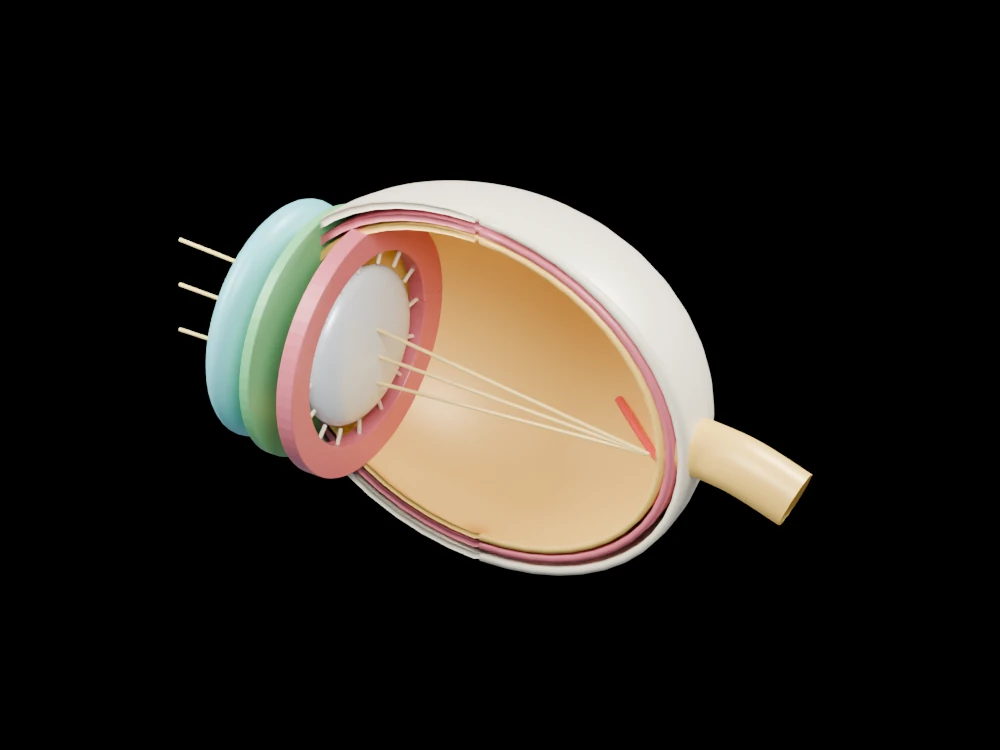 An artificial retina turns near-infrared light into signals in mice
 Engineered cartilage grafts show promise in a 17-patient study
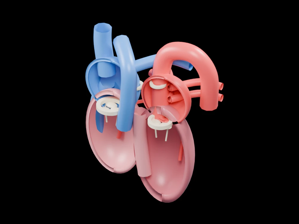 Lab-grown heart cells reach an early human trial: what did it show?
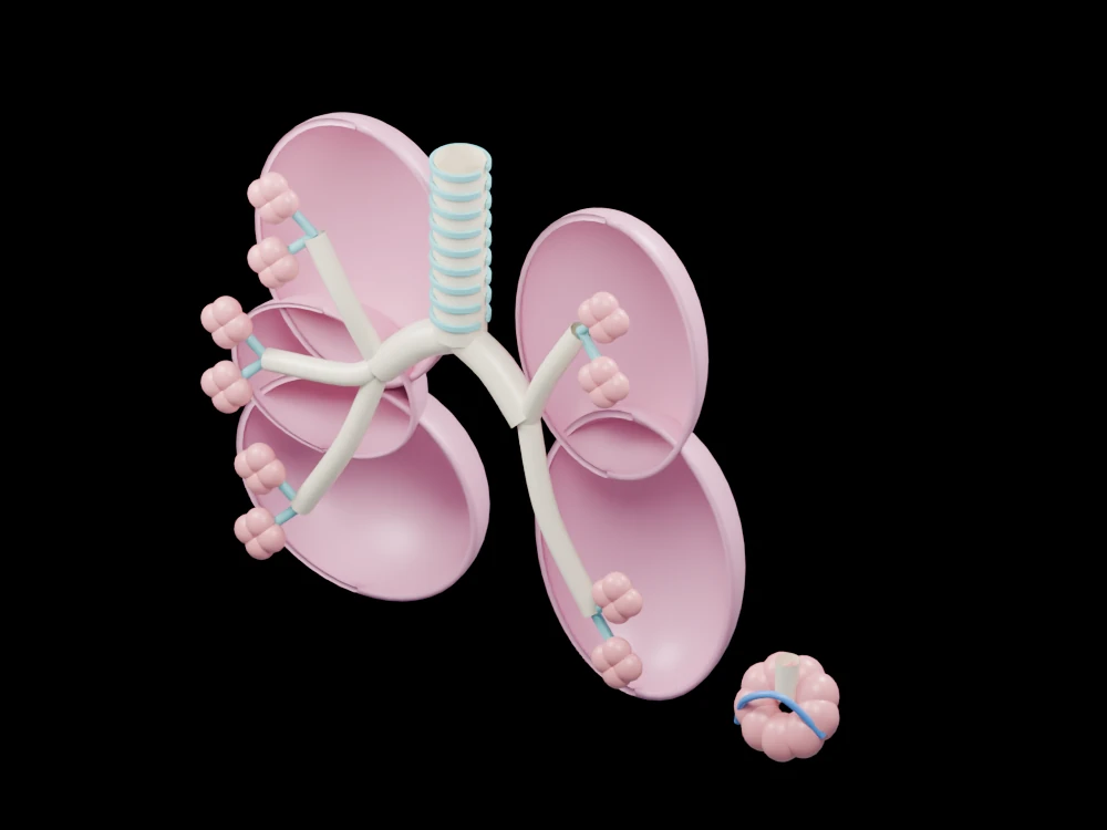 Lung repair crosses an anatomical boundary in a mouse study
 A pig-kidney transplant exposes an early immune hurdle
 AI maps 36 million cells into a shared biological reference
 Evo 2 reads a million DNA letters at once: why that matters
 A flexible protein gate may explain traffic inside mitochondria