

Biological research · Research insights
A flexible protein gate may explain traffic inside mitochondria
An integrated structural model examines how Mic60–Mic19 could regulate passage at crista junctions. Discover why membrane shape and molecular movement belong together.
The folds inside a mitochondrion are often described as extra membrane area. Their connections also matter. Narrow junctions link cristae to the surrounding inner-membrane system, creating places where structure can influence the movement of proteins, metabolites and ions.
Latest breakthrough
A September 2026 Nature Communications paper investigates the human Mic60–Mic19 subcomplex at mitochondrial crista junctions. It combines a crystal structure of part of Mic60 with prior structural information, molecular-dynamics modelling and cross-linking evidence. The resulting model examines how a flexible protein assembly can span the junction and behave as a diffusion barrier. This is an integrative structural-biology result: experimental constraints and computation work together. It is not a direct video of every molecule passing through a living mitochondrion, nor a demonstrated treatment for mitochondrial disease.
Nathanail and colleagues. Nature Communications (2026). Read the original study.
How the method works
The researchers integrated a crystal structure of part of Mic60 with other structural information, molecular-dynamics modelling and cross-linking evidence. A crystal structure constrains an arrangement of atoms; cross-linking supplies evidence about proximity; molecular dynamics explores possible motion within a physical model. Combining them can narrow the plausible arrangements of a flexible assembly. None of those methods alone is a recording of all traffic through a living crista junction. The resulting barrier model therefore needs to be understood as an evidence-constrained explanation of structure and possible function.
A simple analogy
Several clues can help reconstruct a flexible doorway: a detailed drawing of one hinge, measurements of which parts sit nearby and a simulation of how it moves. Together they are more informative than one clue alone. A mitochondrial junction is molecular and fluctuating, so the doorway picture should not be mistaken for a rigid gate opening on command.
A fold can create a distinct environment
When a membrane bends into a narrow-connected compartment, movement into and out of that region can become constrained. Proteins positioned at the connection may further influence that exchange. The consequence is that two regions of one continuous membrane need not behave as a perfectly mixed space. Geometry and molecular organisation can cooperate to maintain local conditions.
Why combining methods is useful
Our interpretation is that flexible assemblies are a good example of why one measurement technique may be insufficient. A partial structure supplies detail, while other experiments constrain how components fit together. Simulation can then test the behaviour of a proposed arrangement. The value lies in agreement among these kinds of evidence, with assumptions kept visible.
From a plausible barrier to broader biology
The model's interpretation needs to remain tied to the structural and biochemical evidence used to build it. Different cellular conditions may change the assembly or the surrounding membrane. Further experiments can test its functional consequences. The possible societal value is improved understanding of cellular energy organisation, while specific diagnostic or therapeutic applications require separate evidence. This local structural question complements large-scale comparison of cell identities, whose data representations do not resolve every organelle mechanism.
Inspect the folds and their connections
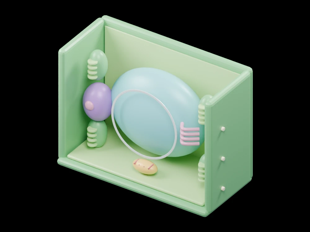
Open the mitochondrion cutaway and distinguish the outer membrane, inner membrane, crista folds and matrix. Pay attention to the regions where folds join the surrounding inner boundary. The gallery depicts organelle organisation at a teaching scale. It does not resolve the Mic60–Mic19 complex or compute diffusion, but it makes the location of the research question much easier to visualise.
Related articles
 AI maps 36 million cells into a shared biological reference
 Lab-grown heart cells reach an early human trial: what did it show?
 Evo 2 reads a million DNA letters at once: why that matters
 Lung repair crosses an anatomical boundary in a mouse study
 A pig-kidney transplant exposes an early immune hurdle
 Spinal stimulation strengthens arm movement in a small stroke study
 A three-person study explores a new angle on recurring UTIs
 Rubisco changes improve photosynthesis in experimental plants
 A protein helps explain how developing leaf veins find direction